
Cone beam computed tomography (CBCT) gives you a clear, three-dimensional look at your teeth, jaws and surrounding structures in a single, comfortable scan. Instead of relying on flat images, our team uses detailed 3D data to plan care with accuracy and explain findings in everyday language. You see what we see, which helps you feel informed and confident at every step at Michael K. Bassiri DDS.
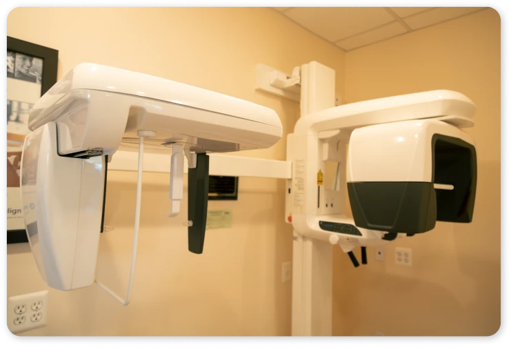
CBCT Imaging: Precise Views for Confident Care
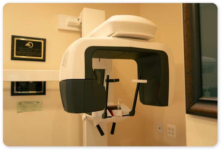
Our dentist uses a compact CBCT unit that rotates gently around your head while you stand or sit. The scan typically lasts less than a minute and captures precise images with focused exposure. Because the technology targets a small area, your dentist can study critical details without subjecting you to lengthy imaging sessions. Dr. Michael Bassiri selected this system to improve clarity while keeping your visit efficient and comfortable.
Why CBCT Matters
Traditional images are helpful, but they can hide depth and fine detail. CBCT in Fairfax, VA, adds dimension, so your dentist can evaluate tooth position, bone contours, joint spaces and airway shape from multiple angles. This level of insight supports safer decisions and smoother appointments. It also allows clearer conversations, since we can rotate and zoom the scan chairside to answer your questions in real time.
Benefits You Can Feel and See
- Precise 3D visuals that reduce uncertainty
- Focused exposure designed for dental use
- Fast, comfortable scan with no closed tunnel
- Clear explanations with on-screen education
- Streamlined planning that saves time
- Secure digital files for easy coordination
What to Expect During Your Scan
You will remove glasses or metal accessories, then step into position. The unit will rotate quietly while you remain still. The process is quick and noninvasive. After the scan, your dentist will review the 3D images with you, highlight areas of interest and outline next steps in simple terms. If you prefer, we offer bilingual explanations to ensure you understand every option.
How We Use CBCT Information
CBCT helps your dentist assess:
- Tooth alignment and root orientation
- Jaw joint health and bite relationships
- Airway space and sinus proximity
- Bone quality and volume for planning
- Cysts, lesions or other unexpected findings
This comprehensive picture reduces guesswork and supports a plan tailored to your goals, health history and comfort level. Because files are digital, we can share them securely when collaboration is needed, which helps keep your timeline on track.
A Modern, Patient-Centered Experience
From scheduling to follow-up, our focus is respect, clarity and comfort. We keep wait times short, maintain a calm environment and use technology that makes appointments smoother. You will never feel rushed, and your questions will always guide the pace of our discussion.
Schedule Your Visit
Ready to experience clear imaging and clear answers with CBCT imaging in Fairfax, Virginia? Call 703-385-9161 to reserve a visit. Our dentist will walk you through the scan, review your images side by side and help you choose the path that fits your needs. High-definition insight and a friendly, modern approach await.
FAQs About CBCT Imaging
Does CBCT use more radiation than standard dental X-rays?
CBCT uses a cone-shaped beam and a limited field of view designed for dentistry. Dose depends on the area scanned and settings, but it is typically lower than a hospital-grade medical CT and higher than a single 2D X-ray. Your dentist selects the smallest scan needed to answer a specific question.
Who should avoid CBCT imaging?
Tell the team if you are pregnant or think you may be pregnant. Extra caution is taken and nonurgent scans are often postponed. Patients with neck mobility limits or difficulty standing can be accommodated with seated positioning and stabilization to keep the scan brief and still.
Will dental work or metal jewelry affect the images?
Metal can create streaks or scatter on any 3D scan. You will be asked to remove earrings, nose rings and removable partials when possible. Fixed dental work is common and usually manageable. Your dentist can adjust the field of view and angulation to minimize artifacts.
How are results delivered and who reviews them?
Your images are generated immediately, then reviewed by your dentist and, when appropriate, securely shared with collaborating clinicians. You receive a summary in clear language, and the files remain part of your protected health record for future reference or comparison.